Product Name
Starting at $5000
EPIQ Elite Elevate combines exceptional image quality with intelligent, tailored clinical insights—enabling clinicians to deliver faster, more confident diagnoses for a broader range of patients.
Engineered with advanced diagnostic capabilities, real-time processing power, and premium performance features, EPIQ Elite is built to thrive in today’s most complex and fast-paced healthcare environments. From routine exams to high-acuity cases, it meets the evolving demands of modern care with precision, efficiency, and reliability.
Image Gallery

Product Features

Auto Doppler: Intelligent Automation for Streamlined Vascular Imaging
Auto Doppler on the Philips EPIQ Elite streamlines vascular imaging by automatically optimizing flow sensitivity and spatial resolution in real time.
This intelligent automation reduces the number of manual setup steps from 10 to just 3, significantly accelerating workflow without compromising image quality. By minimizing repetitive button pushes—cut by an average of 68%*—Auto Doppler allows clinicians to focus more on patient care and diagnostic interpretation, not machine operation. The result: faster, more consistent Doppler exams with improved efficiency and reduced exam variability across users and patient types.
PureWave and xMATRIX: Advanced Transducer Technologies for Exceptional Imaging
Experience the power of PureWave for exceptional imaging—even in technically challenging patients
PureWave crystal technology is the most significant advancement in piezoelectric transducer materials in over 40 years. With perfectly uniform crystals, PureWave delivers greater bandwidth and up to twice the efficiency of traditional ceramic materials—resulting in sharper images, deeper penetration, and more diagnostic confidence.


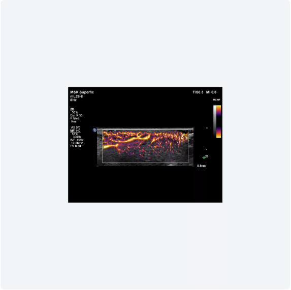
MicroFlow Imaging HD: Enhanced Detection of Low-Velocity Blood Flow
Designed to detect slow and weak blood flow in tissue, MicroFlow Imaging (MFI) overcomes many limitations of conventional Doppler techniques
This proprietary technology reveals small vessel blood flow with high resolution and minimal artifacts—while maintaining excellent frame rates and preserving 2D image quality. Advanced artifact reduction techniques ensure accurate visualization of microvascular anatomy, supporting more confident clinical assessments.
XRES Pro: Next-Generation Image Processing for Exceptional Clarity
At real-time frame rates, XRES Pro applies multi-parametric precision filters that subdivide and analyze image data—sharpening borders, enhancing interfaces, and delivering exceptional tissue conspicuity
This advanced technology also improves assessment of plaque morphology and offers full user adjustability, allowing clinicians to tailor enhancement levels to specific imaging needs—elevating diagnostic confidence across a wide range of patients.


xMatrix Transducers, Powerful and Unmatched
No other premium ultrasound system supports xMATRIX—the most advanced suite of ultrasound transducers available today
Capture ultra-thin 2D slices with precision, and leverage Live xPlane imaging to view two full-resolution planes simultaneously—doubling your clinical information without extending scan time. xMATRIX also delivers near-isovoxel resolution, enabling detailed visualization from any angle within the volume for greater diagnostic confidence.
Next Gen AutoSCAN: Real-Time Brightness Optimization for Consistent Image Quality
Philips Next Gen AutoSCAN enhances image uniformity by adaptively adjusting brightness at every pixel in real time—minimizing the need for manual adjustments and improving transducer plunkability.
With pixel-by-pixel optimization, it reduces button pushes by up to 54%, streamlining workflows and delivering consistent, high-quality imaging with less effort.


24” HD MAX Display: Immersive Clarity with True-to-Life Color Accuracy
This next-generation 24” HD MAX monitor delivers an exceptional ultrasound viewing experience, featuring a 10-bit ultra-wide color gamut capable of displaying billions of colors for true-to-life color accuracy.
With high-contrast dynamic range and enhanced black levels, it allows for subtle differentiation of grayscale values—critical for detailed imaging. The display also offers superior off-angle visibility, ensuring consistent image quality and diagnostic clarity from anywhere in the scanning room.
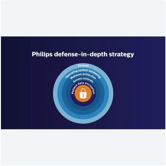
Powerful System Security: Protecting Sensitive Patient Data
As healthcare organizations face increasing cyber threats, data protection has become a critical priority—driving projected cybersecurity spending beyond $65 billion over the next five years. With ultrasound systems often operating in both wired and wireless environments, security is essential.
That’s why Philips has made system protection a core focus. The EPIQ Elite platform is built on a secure Windows 10 operating system and designed with a robust, multi-layered defense-in-depth architecture. It delivers five integrated layers of protection to help safeguard patient data, ensure compliance, and maintain confidence in every clinical setting.
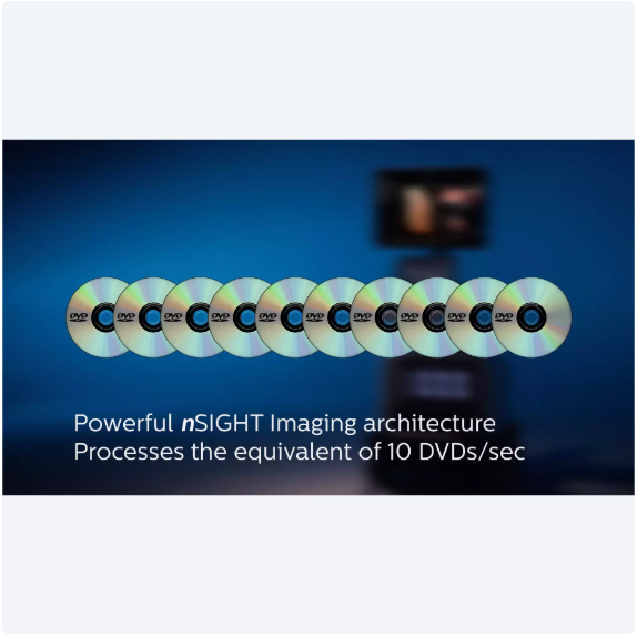
nSIGHT Imaging: Redefining Ultrasound Clarity and Performance
nSIGHT Imaging goes far beyond the limits of conventional ultrasound—delivering remarkable definition, clarity, and real-time performance.
At its core is a proprietary architecture that combines a custom multi-stage precision beamformer with massive parallel processing (MPP). This technology captures a vast amount of acoustic data from every transmit operation and reconstructs it digitally with mathematically optimized focal processing. The result: extraordinary image uniformity, enhanced penetration, and exceptional frame rates—all working together to support faster, more confident clinical decisions.
Super Resolution MVI and Time of Arrival: Advancing CEUS with Greater Precision
Philips Super Resolution MVI for contrast-enhanced ultrasound (CEUS) builds on the legacy of Contrast MVI with next-generation super-resolution processing and advanced motion compensation—delivering up to a 200% improvement in spatial resolution. This breakthrough allows clinicians to visualize microvascular flow with remarkable clarity, even in challenging scenarios.
The integrated Time of Arrival (TOA) map further enhances diagnostic insight by visualizing the relative time microbubbles enter points of interest within the imaging plane, supporting precise perfusion assessment and improved interpretation of contrast dynamics.


Flow Viewer: Enhanced Visualization of Cardiac and Vascular Architecture
Flow Viewer is Philips’ advanced color flow enhancement tool, designed to deliver a 3D-like rendering of vascular and fetal cardiac anatomy.
By adding depth and dimension to flow imaging data, Flow Viewer enhances both the diagnostic clarity and visual appeal of ultrasound scans. This feature is compatible across all color imaging modes—including CFM, CPA, CPAd, MFI, and MFI HD—allowing clinicians to better assess structural relationships and blood flow dynamics in real time.
CEUS High Frame Linear and Auto Scan: Enhanced Contrast Imaging for Thyroid and Beyond
Achieve a 67% increase in frame rate and a 76% expanded field of view when using the Philips eL18-4 transducer for CEUS thyroid imaging***. These significant performance gains provide clearer, more comprehensive views of vascular dynamics in real time.
In addition, CEUS Auto Scan enhances image uniformity and sensitivity by automatically optimizing brightness and gain—reducing manual adjustments and improving consistency across scans.


Auto Segmental Wall Motion Scoring: Objective LV Assessment at a Glance
Auto SWMS delivers automated evaluation of left ventricular wall motion using a standardized 17-segment bullseye display.
This advanced tool enhances the objectivity and consistency of LV assessments, helping clinicians achieve greater reproducibility, efficiency, and confidence in cardiac workflows—particularly in high-volume or time-sensitive environments.
AutoStrain LV: Automated EF and Mid-Layer Strain for Efficient, Reproducible LV Assessment
The latest enhancements to AutoStrain deliver fast, consistent results as part of a comprehensive left ventricular (LV) assessment—all within a single, streamlined application. Integrated AI technology, including Smart View Select, automatically identifies and selects optimal 2D images for LV analysis, reducing manual effort and increasing reproducibility.
With automated ejection fraction (EF) and mid-layer strain measurements, AutoStrain improves workflow efficiency while supporting confident clinical decisions.


Contrast-Enhanced Ultrasound (CEUS): Real-Time, Radiation-Free Diagnostic Insight
CEUS expands the clinical power of ultrasound—particularly in liver imaging—by enabling real-time assessment of enhancement patterns in suspicious liver lesions. It also offers a non-ionizing alternative for evaluating vesicoureteral reflux in pediatric patients.
With CEUS, clinicians gain dynamic, contrast-based imaging without the risks associated with radiation exposure, enhancing diagnostic confidence while supporting patient safety across a range of applications.
Auto ElastQ: Next-Generation Automated Liver Elastography
Auto ElastQ delivers fast, reliable liver stiffness measurements through real-time, quantitative shear wave elastography—streamlining liver health assessments with minimal user interaction.
Designed to simplify workflow and enhance reproducibility, Auto ElastQ provides automated region-of-interest placement and immediate results, supporting early detection, staging, and ongoing monitoring of liver disease with greater clinical confidence.


FlexVue with Orthogonal View: Effortless Access to Challenging Anatomical Planes
FlexVue with Orthogonal View provides intuitive tools to extract and visualize difficult-to-obtain anatomical planes from 3D data sets with ease. This advanced imaging capability enhances diagnostic precision by offering flexible plane acquisition and seamless navigation through complex anatomy.
Paired with a robust measurement package, it enables accurate quantification and supports confident decision-making across a variety of clinical applications.
Image Fusion and Navigation: Integrated Guidance for Confident Clinical Decisions
Simplify complex diagnostic and interventional procedures with fully integrated image fusion and navigation capabilities—directly on the ultrasound system. Philips' fusion technology enables fast, accurate alignment of CT, MR, or PET with live ultrasound imaging, streamlining workflows and enhancing diagnostic confidence, even in the most challenging cases.
With support for a wide range of transducers—including the X6-1 xMATRIX, C5-1, C9-2, eL18-4, L12-5, C10-4ec, S5-1, and the new mC7-2—you can expand fusion and navigation across multiple clinical applications, all while improving efficiency and precision at the point of care.

Anatomically Intelligent Ultrasound: AI-Driven Precision for Faster, More Reproducible Exams
At the core of the EPIQ Elite platform is Philips-exclusive Anatomical Intelligence Ultrasound (AIUS), a next-generation technology that transforms ultrasound from a passive imaging tool into an actively adaptive diagnostic partner. By leveraging advanced organ modeling, automated image slicing, and intelligent quantification, AIUS simplifies exam workflows, enhances reproducibility, and delivers deeper clinical insight.
AIUS powers key features like HeartModel, AI Breast, and Auto-Registration for Image Fusion and Navigation, helping clinicians perform complex exams with greater speed, consistency, and confidence.
Elastography: Comprehensive Tissue Stiffness Assessment with Strain and Shear Wave Imaging
The EPIQ Elite platform delivers advanced elastography capabilities, supporting both strain and shear wave imaging techniques for a comprehensive assessment of tissue stiffness. Highly sensitive strain imaging enables rapid, qualitative evaluation across a wide range of clinical applications.
ElastQ Imaging, Philips’ proprietary shear wave elastography solution, uses a unique pulsing strategy to generate and measure shear wave propagation speed—providing real-time, quantitative stiffness measurements. A built-in confidence map helps ensure measurement accuracy by guiding clinicians to regions with optimal shear wave quality, supporting consistent, reproducible results.
Disclaimer
Lorem Ipsum is simply dummy text of the printing and typesetting industry. Lorem Ipsum has been the industry's standard dummy text ever since the 1500s, when an unknown printer took a galley of type and scrambled it to make a type specimen book.
Specifications
This is paragraph text. Click it or hit the Manage Text button to change the font, color, size, format, and more. To set up site-wide paragraph and title styles, go to Site Theme.
Documentation
Product Video
FAQS
Frequently Asked Questions
What distinguishes the EPIQ Elite Elevate from other ultrasound systems?
The EPIQ Elite Elevate is a premium ultrasound platform designed to deliver exceptional image quality and advanced diagnostic tools. It features AI-assisted workflows, a high-resolution 24" HD MAX display, and supports a wide range of clinical applications, including cardiology, radiology, obstetrics, and point-of-care settings.
How does AI integration enhance the EPIQ Elite Elevate's performance?
The system incorporates Anatomical Intelligence Ultrasound (AIUS), which utilizes advanced organ modeling and image slicing to facilitate faster and more reproducible analyses. Features like AutoStrain and Auto ElastQ automate measurements, reducing exam times and improving diagnostic confidence.
What are the key imaging technologies used in the EPIQ Elite Elevate?
- The EPIQ Elite Elevate employs several advanced imaging technologies:
- nSIGHT Imaging: Enhances image quality through advanced beamforming and image reconstruction.
- PureWave Crystal Technology: Provides uniform, high-quality imaging across various patient types.
- xMatrix Transducers: Allow for quick and easy volume acquisition, supporting multiple imaging modes in a single transducer.
Which clinical applications are supported by the EPIQ Elite Elevate?
The system is versatile and supports a broad spectrum of clinical applications, including:
- Cardiology: Advanced cardiac imaging capabilities, including 3D and 4D echocardiography.
- Obstetrics and Gynecology (OB/GYN): High-resolution imaging for fetal and maternal health monitoring.
- Radiology: Comprehensive imaging for abdominal, vascular, musculoskeletal, and small parts.
- Point-of-Care: Rapid, reliable imaging suitable for emergency and critical care settings.
How does the EPIQ Elite Elevate improve workflow efficiency?
The system is designed with features that streamline workflows:
Quick Launch Presets: Reduce image optimization time by up to 50%.
Auto ElastQ: Automates liver elastography, cutting exam times by up to 60%.
- Intuitive Interface: Simplifies operation, reducing the learning curve for new users.
What display features enhance the user experience?
The EPIQ Elite Elevate features a 24" HD MAX display with:
- 10-bit Color Depth: Provides accurate color reproduction with billions of colors.
- High-Contrast Dynamic Range: Enhances grayscale delineation for subtle image details.
- Wide Viewing Angles: Ensures clear visualization from various positions in the scanning room.
Is the EPIQ Elite Elevate compatible with existing hospital systems?
Yes, the system supports DICOM and HL7 standards, ensuring seamless integration with hospital information systems. It also offers remote access capabilities, allowing clinicians to share and review images and data remotely.