
Checklist for 2026: 12 Steps From Delivery to Go-Live
Purchasing the right OB/GYN ultrasound machine is an important milestone. It is not the end of the equipment-planning process.
Between signing the proposal and scanning the first scheduled patient, a practice may need to coordinate delivery, room preparation, network connectivity, transducers, software, presets, reporting, applications training, quality checks, staff schedules, and the transition from existing equipment.
When those steps are handled separately—or left until installation day—a technically successful equipment purchase can still create an unnecessarily difficult launch.
For a private OB/GYN practice, maternal-fetal medicine program, fertility clinic, women’s imaging center, or hospital department, the objective should be straightforward: the new ultrasound system should arrive in an environment that is ready for it, connect to the systems it needs, support the intended examination mix, and give users enough preparation to begin working confidently.
This 2026 installation checklist provides a practical framework for getting there.
Why Ultrasound Installation Planning Should Begin Before Delivery
Many implementation problems originate before the machine ever reaches the building.
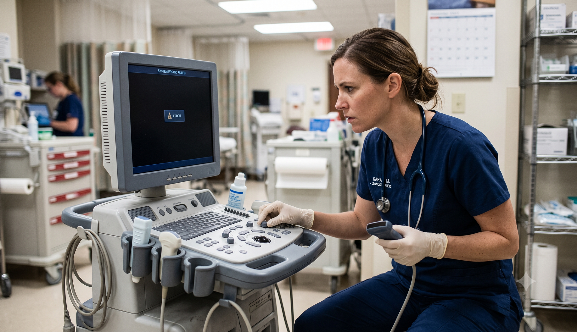
The room may not have the right network connection. A required transducer may have been omitted from the configuration. The practice’s IT team may not know when testing is scheduled. The reporting system may require information that was never provided to the equipment vendor. Sonographers may be booked with patients during the only available training session.
None of these problems necessarily means the practice selected the wrong ultrasound machine.
They mean installation was treated as delivery rather than implementation.
A better approach is to make the installation plan part of the purchasing process. Preferred Medical’s current buyer’s guide already recommends confirming connectivity, installation responsibilities, training, service, and proposed configuration before approval. The installation phase is where those requirements become operational.
1. Assign One Implementation Lead
Every installation should have a clearly identified internal coordinator.
That person does not need to be the clinical expert, IT expert, and equipment expert simultaneously. The role is to make sure the appropriate people are involved and that open items have owners.
Depending on the organization, the implementation team may include:
- Practice administrator or operations leader
- Physician champion
- Lead sonographer
- Ultrasound or imaging manager
- Information technology representative
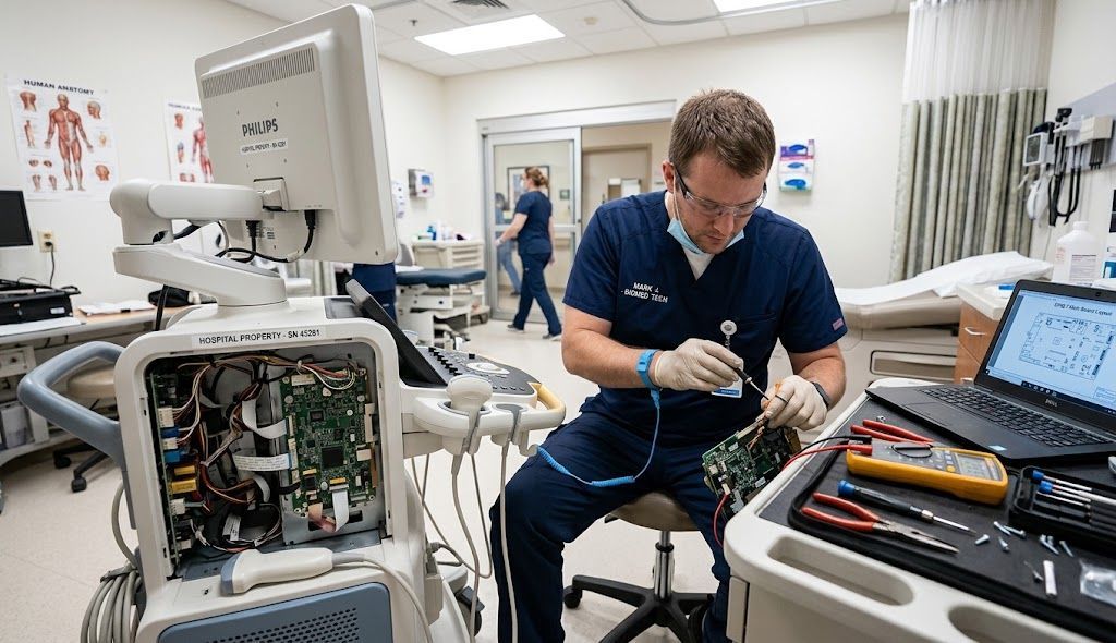
- Biomedical or healthcare technology management personnel
- PACS or reporting-system representative
- Equipment vendor
- Clinical applications specialist
- Field-service personnel
- Facilities or construction staff
- Infection-prevention personnel when appropriate
For a smaller private practice, several of these responsibilities may belong to the same person.
Create one shared implementation document containing the equipment configuration, delivery date, training date, IT requirements, room-readiness tasks, key contacts, and unresolved questions.
2. Confirm the Exact Equipment Configuration
Before preparing the room, confirm exactly what is arriving.
The final configuration should identify:
- Manufacturer and model
- New, demonstration, or pre-owned condition
- Software version
- Installed clinical packages
- Measurement packages
- 3D/4D capabilities when applicable
- Automation features
- Number and type of transducers
- Printers or peripherals
- Footswitches
- Gel warmers or accessories
- Batteries for applicable compact systems
- DICOM or connectivity options
- Reporting interfaces
- Warranty information
- Service coverage
- Included applications training
For OB/GYN imaging, pay particular attention to the transducer package. A system can arrive fully operational and still be clinically incomplete if the probes required for the practice’s examination mix were not included.
Compare the delivered configuration with the final approved proposal rather than relying on memory from the demonstration.
3. Prepare the Ultrasound Room
Room readiness affects workflow long after installation day.
Measure the proposed location with the ultrasound system, examination table, sonographer chair, patient access, cabinets, computer workstation, printer, storage, doors, and frequently used supplies in mind.
Evaluate:
- Electrical access
- Network connections
- Equipment footprint
- Monitor movement
- Control-panel movement
- Sonographer positioning
- Patient-table positioning
- Chair clearance
- Probe and cable management
- Storage access
- Lighting
- Cleaning access
- Ventilation requirements
- Movement of the system through doorways
- Space needed for physicians or additional staff
Ergonomics deserves particular attention. The Society of Diagnostic Medical Sonography recommends providing adequate scanning-room space so patient positioning, workstation positioning, and ultrasound equipment can be optimized.
Do not evaluate room size only by asking whether the machine physically fits.
The better question is whether the people using the machine can position themselves, the patient, the table, the monitor, and the control panel appropriately throughout a normal schedule.
4. Plan the Delivery Route
A cart-based ultrasound system may need to pass through loading areas, elevators, corridors, security checkpoints, and clinical spaces before reaching its room.
Before delivery, confirm:
- Shipping address and receiving contact
- Building access hours
- Loading-dock availability
- Elevator dimensions and restrictions
- Doorway clearance
- Stairs or other access limitations
- Parking requirements
- Security procedures
- Packaging-removal responsibilities
- Whether the old system must be moved
- Who will accept delivery
For a new office or construction project, verify that the planned installation date occurs after the room is genuinely ready—not simply after the contractor’s original completion estimate.
5. Complete the IT and Connectivity Plan Before Installation Day
Modern ultrasound systems often operate within a broader information environment rather than as isolated devices.
Depending on the practice, the system may need to communicate with:
- PACS
- Modality worklist
- Reporting software
- Electronic health record systems
- Image archives
- Network storage
- Remote-support systems
- Enterprise authentication systems
DICOM Modality Worklist, for example, is designed to provide an imaging acquisition device with scheduled procedure information.
Before installation, determine who is responsible for:
- Network addressing
- DICOM configuration
- AE titles and ports
- Worklist setup
- PACS destinations
- Reporting integration
- User authentication
- Firewall changes
- Remote-support access
- Storage configuration
- Testing patient-data flow
- Testing images and clips
- Confirming timestamps and identifiers
- Resolving rejected studies
Cybersecurity belongs in this discussion as well. FDA guidance describes medical-device manufacturers and healthcare delivery organizations as sharing responsibility for appropriate cybersecurity safeguards, while healthcare organizations are responsible for evaluating and protecting their networks.
A practice should therefore involve its IT and security personnel before connection rather than simply allowing the equipment to be placed on the network and addressing questions afterward.
6. Build Clinical Presets Around the Actual Examination Mix
Factory presets are a starting point.
A useful implementation process adapts the system to the examinations, users, documentation requirements, and workflow of the organization.
Depending on the practice, configuration may include presets for:
- First-trimester obstetrics
- Second- and third-trimester obstetrics
- Gynecologic examinations
- Transvaginal imaging
- Pelvic imaging
- Doppler examinations
- Fertility monitoring
- Follicular assessment
- Maternal-fetal medicine
- Fetal cardiac imaging
- 3D/4D acquisition
- Breast imaging
- Procedure guidance
The practice should also determine which measurements, annotations, calculations, image labels, reporting fields, and user preferences need to be standardized.
For multisite groups, consider whether common presets should be established across locations.
The objective is not to eliminate appropriate clinical customization. It is to reduce unnecessary differences that make training, coverage, and workflow harder than they need to be.
7. Verify Transducers and Accessories
Each transducer should be matched against the purchase documentation and intended application.
Record:
- Probe model
- Serial number
- Clinical purpose
- Condition
- Warranty status
- Compatible port
- Storage location
Confirm that the practice has an approved process for cleaning and handling both external and internal-use transducers consistent with manufacturer instructions and organizational infection-control policies.
Also identify what will happen if a critical probe fails.
A functioning console may still be unable to support a key examination if the required endocavitary, curved-array, volume, linear, or specialty transducer is unavailable.
8. Establish an Equipment and Quality Baseline
Installation should create a documented starting point for the new system.
Record the machine’s:
- Model and serial number
- Software revision
- Installed options
- Transducers
- Accessories
- Installation date
- Warranty start date
- Service agreement
- Network configuration where appropriate
- Training completion
- Acceptance or operational testing records
AIUM’s 2026 accreditation standards state that ultrasound equipment should be kept in optimal operating condition and undergo documented quality-assurance testing at least annually, or more frequently if issues arise.
The exact acceptance and QA procedures required at installation depend on organizational policies, accreditation status, manufacturer guidance, applicable regulations, and the clinical environment.
The important operational principle is to begin ownership with a documented baseline rather than trying to reconstruct equipment history years later.
9. Schedule Applications Training Around Real Users
Training should not be an afterthought attached to the delivery calendar.
The people who will actually use the system should be available.
A practical training agenda may cover:
- System startup and shutdown
- User profiles
- Transducer selection
- Image optimization
- Clinical presets
- Measurements
- Calculations
- Doppler controls
- 3D/4D tools when applicable
- Automation functions
- Image review
- Reporting workflow
- Worklist use
- Image transfer
- Data management
- User customization
- Examination-room ergonomics
- Troubleshooting basics
Preferred Medical currently states that customized applications training is included with the systems it sells and can be delivered on-site or virtually, with training tailored to the equipment, specialty, workflow, and team.
For a busy practice, it may be more effective to divide education into stages rather than attempting to teach every advanced feature on day one.
Initial training can focus on safe, efficient routine workflow. Follow-up sessions can address optimization and advanced applications after users have gained practical experience.
10. Test the Entire Workflow—Not Just the Image
A technically excellent ultrasound image does not prove that implementation is complete.
Before full go-live, test a representative workflow from beginning to end.
For example:
- Create or select the scheduled examination.
- Verify patient information on the ultrasound system.
- Select the appropriate preset.
- Acquire test images.
- Perform required measurements.
- Save images and clips.
- Send the study to the intended archive.
- Confirm receipt.
- Confirm reporting workflow.
- Verify that required identifiers and documentation appear correctly.
Include the people who normally work at each point in that process.
A workflow issue discovered during a controlled test is considerably easier to address than one discovered during a full patient schedule.
11. Plan the Go-Live Schedule Conservatively
Avoid treating the first clinical day on a new platform as an ordinary full-volume day when operationally possible.
Users may need additional time to:
- Locate controls
- Adjust presets
- Learn measurements
- Navigate worklists
- Change transducers
- Complete documentation
- Ask applications questions
Consider temporarily reducing the first-day schedule, placing experienced users in the room, or having additional applications support available.
If the practice is replacing an older machine, keep the existing system available until the new platform has been installed, connected, tested, and accepted whenever practical.
The most difficult time to discover a network, configuration, or workflow problem is after the previous equipment has already left the building.
12. Schedule a Post-Installation Optimization Review
Go-live is not the final implementation milestone.
After users have completed enough examinations to understand the new workflow, collect structured feedback.
Ask:
- Which presets need refinement?
- Are measurements arranged efficiently?
- Are users taking unnecessary steps?
- Is the worklist reliable?
- Are images consistently reaching the archive?
- Are there reporting problems?
- Are transducer assignments practical?
- Are users comfortable with ergonomics?
- Which advanced features require additional training?
- Are there support issues that should be documented?
This review allows the practice to distinguish between temporary learning-curve issues and configuration problems that genuinely need correction.
A Practical OB/GYN Ultrasound Installation Checklist
Before declaring the installation complete, confirm that:
- The delivered system matches the approved configuration.
- Every required transducer is present.
- The room supports appropriate positioning and workflow.
- Electrical and networking requirements are complete.
- PACS, worklist, reporting, or EHR connections have been tested.
- Required cybersecurity review is complete.
- Clinical presets reflect the intended examination mix.
- User accounts and preferences are configured.
- Equipment records have been created.
- Applications training has been completed.
- End-to-end workflow has been tested.
- Service and support contacts are documented.
- The transition from the previous equipment has been planned.
- Follow-up optimization is scheduled.
Frequently Asked Questions
How long does it take to install an OB/GYN ultrasound machine?
The physical setup of an ultrasound system may be relatively straightforward, but the complete implementation timeline depends on configuration, delivery logistics, network integration, reporting systems, room readiness, training, testing, and organizational requirements.
Practices should plan around the complete implementation—not simply the time required to move the machine into the room.
Who should be involved in an ultrasound installation?
At minimum, involve the clinical users and the person responsible for practice operations. Organizations with connected imaging environments should also involve IT, PACS or reporting personnel, and biomedical or healthcare technology management staff when applicable.
Should applications training occur on installation day?
It can, but the schedule should provide enough uninterrupted time for meaningful education. For more advanced platforms, staged training or follow-up optimization may provide greater value than trying to cover every feature during one session.
What should be tested before the first patient?
Test the equipment configuration, required probes, clinical presets, measurements, user access, worklist, image transfer, archive or PACS connection, reporting workflow, and any other functions the practice needs during a typical examination.
Should the old ultrasound system be removed before the new one arrives?
When operationally possible, it can be beneficial to keep the previous machine available until the replacement has been installed, tested, connected, and accepted. Space, trade-in terms, data-management requirements, and facility policies may affect the transition plan.
Turn an Equipment Purchase Into a Successful Imaging Launch
The right ultrasound machine matters. So does everything that happens between equipment selection and the first successful clinical day.
Preferred Medical works with OB/GYN practices, maternal-fetal medicine programs, fertility clinics, imaging centers, hospitals, and women’s health organizations to coordinate ultrasound technology planning, configuration, installation, applications training, service, and ongoing optimization.
Preferred Medical currently offers Philips, Mindray, and Alpinion ultrasound solutions and describes its support model as including installation assistance, customized applications training, field service, parts and probe support, and consultative equipment planning.
Already selected a system—or preparing to replace one?
Request an ultrasound implementation consultation with Preferred Medical to review your equipment configuration, room, transducers, connectivity, training needs, service plan, and desired go-live schedule before installation day.
Recommended Articles

Should You Buy, Finance, or Lease an OB/GYN Ultrasound Machine in 2026? A Practice Acquisition Guide

How Many Scans Does It Take to Pay for an OB/GYN Ultrasound Machine? A 2026 ROI and Break-Even Guide