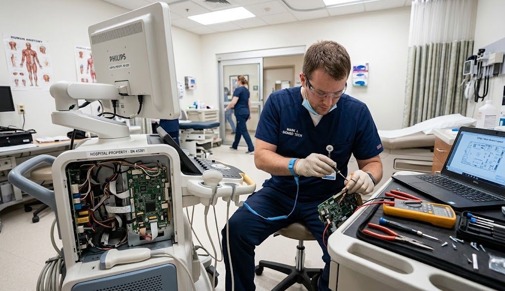

How to Evaluate an OB/GYN Ultrasound Machine Demo in 2026: A Practical Guide for Women's Health Practices
Why an Ultrasound Demonstration Is One of the Most Important Steps in Equipment Selection
Choosing an ultrasound system is a significant decision for any women’s health organization. The equipment must support clinical expectations, fit the daily workflow, accommodate the sonographers who use it, and remain productive for years after installation.
Yet many ultrasound purchasing decisions begin with specifications, product brochures, and quoted prices.
Those resources are important, but they cannot fully demonstrate how a machine will perform inside an actual OB/GYN practice.
A thoughtfully structured OB/GYN ultrasound machine demo helps bridge that gap.
For physicians, sonographers, imaging directors, and practice administrators, a demonstration provides an opportunity to evaluate not simply what a system can do, but how effectively it performs the work required by the organization.
That distinction matters.
An ultrasound system may produce impressive images during a manufacturer presentation but still require extensive manual adjustments during routine examinations. Another may offer sophisticated features that rarely contribute to the practice’s everyday workflow.
In 2026, evaluating ultrasound equipment means examining clinical capability, workflow efficiency, automation, connectivity, training, ergonomics, and long-term support together.
Preferred Medical encourages women’s health organizations to approach demonstrations with clearly defined clinical and operational objectives.
The goal is not to identify the machine with the longest feature list. It is to identify the system that best supports the organization’s imaging program.
1. Define the Clinical Requirements Before Scheduling the Demo
An effective equipment demonstration begins before the ultrasound system enters the examination room.
Practice leaders should identify the examinations performed today, the capabilities needed in the future, and the operational difficulties associated with existing equipment.
A routine OB/GYN practice may primarily perform early pregnancy examinations, pelvic imaging, routine obstetric examinations, and gynecologic assessments.
A maternal-fetal medicine organization may require more demanding anatomical visualization, specialized examination workflows, and advanced imaging capabilities.
Fertility centers may focus more heavily on endovaginal imaging, follicular monitoring, uterine assessment, and efficient repeated measurements.
Multispecialty women’s imaging centers may also require high-frequency linear imaging for breast or superficial examinations.
Because these clinical environments differ, a single generic demonstration is unlikely to address every buyer’s needs.
Before the appointment, create a written list of essential examinations and relevant clinical challenges.
Identify which studies account for the greatest volume, which require the most sonographer time, and which imaging applications create the greatest difficulty on current systems.
This establishes a realistic foundation for evaluating performance.
2. Request a Demonstration That Reflects Actual Patient Workflows
Manufacturer demonstrations often emphasize advanced imaging examples and visually impressive capabilities.
These can be useful, but purchasing decisions should also consider routine examinations.
A practice performing predominantly general OB/GYN imaging should spend meaningful demonstration time on the studies its team completes every day.
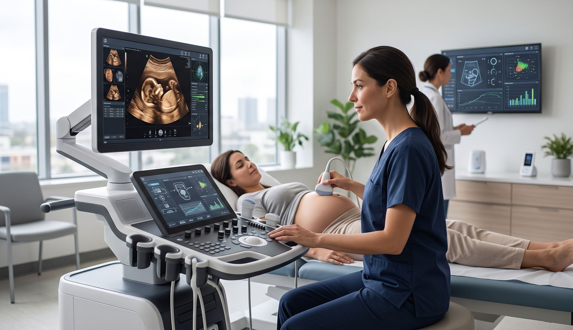
Evaluate the ultrasound machine using representative clinical scenarios and appropriately authorized demonstration subjects or approved test materials.
For obstetric imaging, assess the workflow for routine fetal measurements, anatomical visualization, image optimization, and documentation.
For gynecologic imaging, examine pelvic structures, endovaginal imaging workflow, measurements, and reporting.
For fertility imaging, consider follicular assessment, endometrial evaluation, repeated measurements, and the ability to review prior studies when supported by the configuration.
Demonstrations involving patients should follow applicable consent, privacy, infection-prevention, and organizational policies.
The demonstration should answer a practical question:
Can the system support the examinations our practice actually performs, with the consistency and efficiency our clinicians need?
3. Evaluate Image Quality Across Multiple Clinical Applications
Image quality remains a fundamental part of ultrasound system selection.
However, image quality should not be judged from one exceptionally favorable example.
A meaningful evaluation considers multiple examination types, imaging depths, tissue characteristics, and transducer configurations.
Assess how easily the sonographer can optimize penetration, contrast, spatial detail, and visualization of relevant anatomy.
Examine the image during movement, not only when the system displays a carefully selected still frame.
Observe whether image optimization requires frequent manual intervention.
Modern ultrasound platforms may incorporate processing tools designed to streamline imaging adjustments, but the availability and performance of these tools vary by system and software configuration.
A premium system is not automatically the best operational choice for every practice.
The most relevant measure is whether its imaging capabilities support the clinical work expected from that specific organization.
Clinicians should evaluate diagnostic adequacy using their established professional standards and clinical protocols rather than relying solely on vendor presentation materials.
4. Compare Transducers, Not Just Ultrasound Consoles
One of the most overlooked aspects of an ultrasound demonstration is the transducer package.
Two ultrasound machines from the same manufacturer may perform differently in practice depending on the connected probes and installed applications.
For women's health imaging, common transducer considerations include curved-array probes for abdominal and obstetric imaging, endocavitary probes for pelvic and early pregnancy examinations, volume probes for appropriate 3D/4D applications, and high-frequency linear probes for breast or superficial imaging.
The exact combination depends on clinical requirements.
During the demonstration, verify which transducers are included in the proposed purchase.
Ask whether the system supports additional probes the practice may require as services expand.
Transducer ergonomics should also be evaluated.
Consider probe weight, grip, cable management, cleaning compatibility, and accessibility during scanning.
A transducer that performs well technically but is uncomfortable to use throughout a demanding examination schedule may create operational challenges.
The complete imaging configuration matters more than the console alone.
5. Test Workflow Automation With Realistic Tasks
Artificial intelligence and automated ultrasound measurement technologies are increasingly visible in manufacturer marketing.
These capabilities deserve attention, particularly in practices that perform high volumes of repetitive examinations.
However, the existence of an automated feature does not guarantee meaningful time savings.
A demonstration should evaluate the entire workflow, including image acquisition, automatic measurement, manual correction, confirmation, documentation, and reporting.
For example, Alpinion offers automated imaging functions within its X-CUBE portfolio, including fetal biometry and follicular measurement assistance on supported configurations.
Mindray’s Imagyn I9 incorporates women's health imaging tools and an ergonomically designed interface, while Philips has continued developing software-driven imaging and workflow capabilities for its EPIQ Elite and Affiniti platforms.
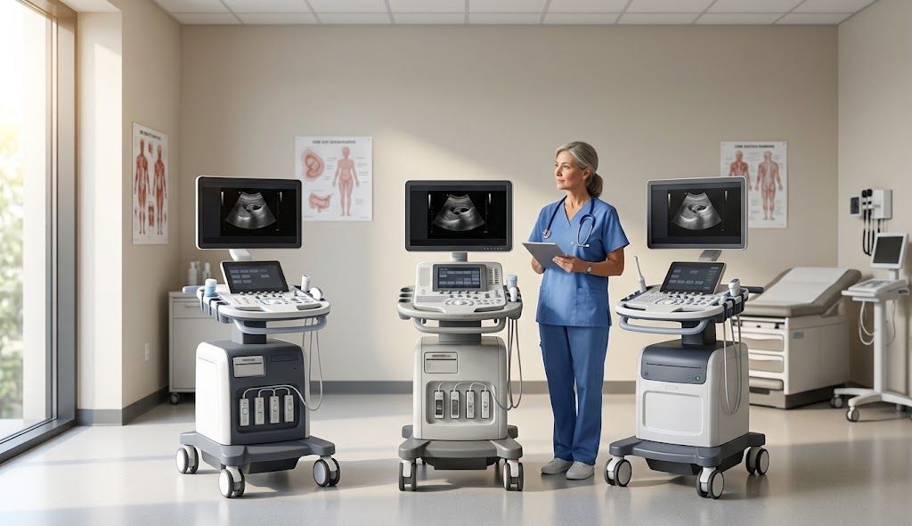
Each platform should be evaluated using the precise software, clinical package, and transducers being quoted.
Automation should assist qualified clinical users, not substitute for their professional judgment.
During a demonstration, consider whether the technology reduces repetitive actions, provides useful measurement assistance, and integrates naturally with the practice’s documentation process.
Ask the demonstrating applications specialist to show what happens when an automatic measurement requires adjustment.
The ability to review, correct, and confirm automated output is just as important as obtaining the initial result.
6. Include Sonographers in the Decision
An ultrasound machine may be selected by an administrator or physician, but sonographers often interact with the system throughout the working day.
Their input should be central to the demonstration process.
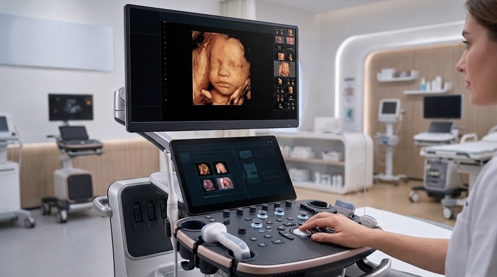
Evaluate the control panel, monitor positioning, touchscreen responsiveness, preset accessibility, transducer connections, and overall physical adjustment options.
- Can the controls be positioned comfortably for different users?
- Can the monitor be viewed without awkward neck positioning?
- Are frequently used functions accessible without unnecessary menu navigation?
- Does the system allow clinicians to move through routine examination steps efficiently?
Ergonomics can influence user comfort, consistency, and workflow.
The Mindray Imagyn I9, for example, features a floating control interface and customizable controls designed with user interaction in mind. Similar ergonomic considerations should be assessed across every manufacturer under consideration.
Instead of asking whether a machine is easy to use, have several team members perform the same representative tasks.
Differences in user experience often become apparent during hands-on interaction.
7. Test Reporting and Connectivity
Image acquisition represents only one part of the ultrasound examination.
The completed study must also be documented, stored, communicated, and made available through the appropriate clinical systems.
During the demonstration, ask the equipment representative to explain available connectivity options.
Depending on the practice’s environment, these may include DICOM image transfer, PACS connectivity, modality worklists, reporting workflows, and integration with applicable electronic medical record systems.
Some functions require additional licensing, interface development, or configuration.
For that reason, buyers should distinguish between functionality demonstrated on a fully equipped machine and what is included in their actual proposal.
Ask how patient information enters the system, how examinations are saved, how images are transferred, and what happens when a network connection is unavailable.
Involve the organization’s information technology team before purchase.
Connectivity should be validated during implementation, not assumed from a product brochure.
8. Evaluate Training Requirements
A new ultrasound system may offer considerable functionality that users will never fully adopt without appropriate training.
Ask what applications training is included with the equipment purchase.
Determine whether training can accommodate multiple clinicians, different experience levels, and varied examination types.
Consider initial instruction, advanced applications training, refresher sessions, and onboarding for new staff members.
A growing practice may also require standardized presets and workflows so different sonographers can produce consistent examination documentation.
The practical value of an ultrasound system depends partly on how confidently the team can use it.
Training should therefore be evaluated as part of the equipment investment rather than treated as an optional afterthought.
9. Ask About Service, Warranty, and Long-Term Support
The equipment demonstration is also an opportunity to assess the support structure behind the technology.
Image quality during a demonstration matters, but so does the ability to keep the system operational.
Ask about preventive maintenance, service response procedures, technical assistance, software support, transducer coverage, and warranty exclusions.
Clarify what happens if a transducer fails or the system requires repair.
Discuss whether loaner equipment or temporary alternatives may be available under the proposed support arrangement.
For organizations with limited ultrasound capacity, even a brief interruption can complicate patient scheduling and staff utilization.
A clear support plan helps practice leaders understand their operational exposure.
Preferred Medical’s consultative approach incorporates equipment planning, training, service, and ownership considerations rather than treating the machine as an isolated purchase.
10. Use a Structured Demonstration Scorecard
After evaluating multiple manufacturers, it can be difficult to remember the differences between machines.
A structured scorecard provides a consistent comparison framework.
Consider assigning each category a rating based on the organization's priorities.
| Evaluation category | Suggested weight |
|---|---|
| Image quality and clinical suitability | 25% |
| Transducers and examination coverage | 20% |
| Sonographer workflow and ergonomics | 15% |
| Automation and reporting | 15% |
| Connectivity and implementation | 10% |
| Training and service support | 10% |
| Expansion flexibility | 5% |
These are illustrative weights, not universal industry standards. Practices should adjust them to reflect their clinical and operational requirements.
Use the same scorecard across demonstrations, document meaningful observations, and identify any features that were not available for testing.
A numerical score can organize discussion, but it should never override essential clinical requirements.
11. Compare the Demonstrated Configuration With the Final Proposal
One of the most important final steps occurs after the demonstration.
Before signing an equipment agreement, confirm that the quoted machine includes the capabilities actually evaluated.
Review the transducer package, software options, automation features, connectivity licenses, accessories, training, installation, and service coverage.
Ask whether demonstrated features are standard, optional, or available only on specific configurations.
This is particularly important when comparing new, demonstration, and pre-owned systems.
The machine that impressed the clinical team may not be identical to the system listed in the final quotation.
A careful proposal review reduces the risk of discovering critical omissions after installation.
Make the Demonstration Part of a Complete Equipment Strategy
An effective ultrasound demonstration should produce more than enthusiasm for new technology.
It should provide a clear understanding of clinical fit, daily usability, implementation requirements, and long-term ownership expectations.
For OB/GYN practices, fertility centers, maternal-fetal medicine programs, and women’s imaging organizations, this approach supports more disciplined equipment decisions.
The best demonstration is the one that answers the organization's most important questions—not necessarily the one with the most impressive presentation.
Take the Next Step With Preferred Medical
Preferred Medical helps women’s health organizations evaluate ultrasound technologies from Philips, Mindray, and Alpinion with attention to clinical requirements, workflow, training, service, and long-term value.
Whether you are replacing an aging system, opening a new ultrasound suite, or comparing multiple platforms, our team can help you develop a practical evaluation strategy.
Contact Preferred Medical to discuss your ultrasound needs and the next steps in selecting the right system for your practice.

Should You Buy, Finance, or Lease an OB/GYN Ultrasound Machine in 2026? A Practice Acquisition Guide

How Many Scans Does It Take to Pay for an OB/GYN Ultrasound Machine? A 2026 ROI and Break-Even Guide