

OB/GYN Ultrasound Glossary: Terms Used by Sonographers and Women's Health Clinicians
The Preferred Medical Team
A plain-language reference to the terms OB/GYN physicians, maternal-fetal medicine specialists, and sonographers use every day, grouped by topic.
Pregnancy Dating and Fetal Biometry
| Term | Definition |
|---|---|
| AC (abdominal circumference) | Measurement around the fetal abdomen at the level of the stomach and umbilical vein. It is the measurement most sensitive to fetal growth problems. |
| BPD (biparietal diameter) | Width of the fetal head measured across the parietal bones. Used with other measurements to estimate gestational age and fetal weight. |
| CRL (crown-rump length) | Length of the embryo or fetus from the top of the head to the bottom of the rump. The most accurate ultrasound measure for dating in the first trimester. |
| EDD (estimated due date) | The projected delivery date, set from the last menstrual period, early ultrasound, or assisted-reproduction dates. Also called EDC. |
| EFW (estimated fetal weight) | Calculated fetal weight based on a formula, commonly Hadlock, combining BPD, HC, AC, and FL. |
| FL (femur length) | Length of the ossified shaft of the fetal femur. A standard biometric measurement. |
| FGR (fetal growth restriction) | A fetus that has not reached its growth potential, usually defined by an EFW or AC below the 10th percentile. Also called IUGR. |
| GA (gestational age) | Age of the pregnancy measured from the first day of the last menstrual period, expressed in weeks and days. |
| GS / MSD (gestational sac / mean sac diameter) | The fluid-filled sac seen early in pregnancy. MSD is the average of its three dimensions. |
| HC (head circumference) | Measurement around the fetal skull in the same plane as the BPD. |
| LGA (large for gestational age) | A fetus or newborn with weight above the 90th percentile for gestational age. |
| LMP (last menstrual period) | First day of the last period, used as the starting point for clinical dating. |
| Macrosomia | Fetal or birth weight above a set threshold, commonly 4,000 or 4,500 grams. |
| SGA (small for gestational age) | A fetus or newborn with weight below the 10th percentile for gestational age. |
| Yolk sac | A small round structure inside the early gestational sac. It is the first structure confirming an intrauterine pregnancy. |
Obstetric Exams, Anatomy, and Findings
| Term | Definition |
|---|---|
| AFI (amniotic fluid index) | Sum of the deepest vertical fluid pockets in four quadrants of the uterus, in centimeters. |
| Anatomy scan | Standard second-trimester survey of fetal anatomy, usually done at about 18–22 weeks. Also called the anatomy survey or level I. |
| Cervical length | Length of the closed cervix, usually measured transvaginally. A short cervix is associated with preterm birth risk. |
| Chorionicity / amnionicity | The number of placentas (chorions) and amniotic sacs in a multiple pregnancy. It is best determined in the first trimester. |
| Detailed anatomy scan | Indication-driven, more comprehensive fetal anatomy exam performed for higher-risk pregnancies. Often called level II, and billed as CPT 76811. |
| Detailed anatomy scan | Indication-driven, more comprehensive fetal anatomy exam performed for higher-risk pregnancies. Often called level II, and billed as CPT 76811. |
| DVP / MVP (deepest or maximum vertical pocket) | Depth of the largest single pocket of amniotic fluid. Used alone or in twins to assess fluid volume. |
| Ectopic pregnancy | A pregnancy implanted outside the uterine cavity, most often in a fallopian tube. |
| FHR (fetal heart rate) | Fetal heartbeats per minute, documented with M-mode or Doppler. |
| Fetal echocardiogram | Detailed evaluation of fetal heart structure and function, usually performed by MFM or pediatric cardiology. |
| Fetal presentation / lie | The part of the fetus closest to the cervix (such as cephalic or breech) and the fetus's orientation relative to the mother's long axis. |
| NT (nuchal translucency) | Fluid space at the back of the fetal neck measured at about 11–14 weeks. Increased NT is associated with chromosomal and cardiac abnormalities. |
| Oligohydramnios / polyhydramnios | Too little or too much amniotic fluid for gestational age. |
| PAS (placenta accreta spectrum) | Abnormally deep attachment of the placenta into or through the uterine wall. Includes accreta, increta, and percreta. |
| Placenta previa | A placenta that covers the internal opening of the cervix. |
| Low-lying placenta | A placental edge within 2 cm of the internal cervical os without covering it. |
| Soft marker | An ultrasound finding that is often normal but slightly raises the likelihood of a chromosomal condition, such as an echogenic intracardiac focus. |
| Three-vessel view / 3VT | Fetal cardiac view showing the pulmonary artery, aorta, and superior vena cava (and trachea in 3VT). Part of the outflow tract assessment. |
| Four-chamber view | Standard transverse view of the fetal heart showing both atria and both ventricles. |
| TTTS (twin-twin transfusion syndrome) | Unbalanced blood flow between monochorionic twins through shared placental vessels. |
| Viability / IUP | Confirmation of a living intrauterine pregnancy (IUP), usually by fetal cardiac activity. |
Doppler and Fetal Surveillance
| Term | Definition |
|---|---|
| AEDF / REDF | Absent or reversed end-diastolic flow in the umbilical artery. A sign of increased placental resistance in growth-restricted fetuses. |
| BPP (biophysical profile) | Scored ultrasound assessment of fetal breathing, movement, tone, and amniotic fluid, often combined with a nonstress test. |
| CPR (cerebroplacental ratio) | MCA pulsatility index divided by umbilical artery pulsatility index. A low ratio can suggest fetal blood-flow redistribution. |
| DV (ductus venosus) Doppler | Waveform from the small fetal vessel connecting the umbilical vein to the heart. Abnormal flow can indicate cardiac strain. |
| MCA (middle cerebral artery) Doppler | Flow measurement in a fetal brain artery. Peak systolic velocity screens for fetal anemia. |
| MCA-PSV | Peak systolic velocity in the middle cerebral artery, expressed in multiples of the median (MoM). |
| PI (pulsatility index) | (Peak systolic − end-diastolic velocity) ÷ mean velocity. A common measure of downstream vascular resistance. |
| RI (resistive index) | (Peak systolic − end-diastolic velocity) ÷ peak systolic velocity. |
| S/D ratio | Peak systolic velocity divided by end-diastolic velocity, often reported for the umbilical artery. |
| UA (umbilical artery) Doppler | Flow assessment in the umbilical cord artery. A core test in monitoring fetal growth restriction. |
| Uterine artery Doppler | Flow measurement in the maternal uterine arteries. Elevated resistance or notching is linked to preeclampsia and placental insufficiency. |
Gynecologic and Fertility Imaging
| Term | Definition |
|---|---|
| Adenomyosis | Endometrial tissue growing into the muscular wall of the uterus, often causing a bulky, heterogeneous uterus. |
| Adnexa | The structures beside the uterus: the ovaries, fallopian tubes, and supporting ligaments. |
| AFC (antral follicle count) | Number of small ovarian follicles, typically 2–10 mm, counted early in the cycle. A marker of ovarian reserve. |
| Corpus luteum | The structure that forms from the follicle after ovulation. It often appears as a cyst with a ring of blood flow. |
| Dominant follicle | The follicle selected to ovulate during a cycle, larger than the others. |
| Endometrial stripe / thickness | The lining of the uterus as seen on ultrasound, measured front to back in the sagittal plane. |
| Fibroid (leiomyoma) | A benign smooth-muscle tumor of the uterus, described by location (submucosal, intramural, subserosal). |
| Follicle monitoring | Serial ultrasound measurement of ovarian follicles during an ovulation induction or IVF cycle. |
| Hydrosalpinx | A fluid-filled, blocked fallopian tube. |
| O-RADS | Ovarian-Adnexal Reporting and Data System, a standardized risk system for describing adnexal masses. |
| OHSS (ovarian hyperstimulation syndrome) | Enlarged ovaries and fluid shifts caused by fertility medications. |
| PCOM (polycystic ovarian morphology) | An increased number of follicles or increased ovarian volume, one criterion for polycystic ovary syndrome (PCOS). |
| Saline infusion sonography (SIS) | Transvaginal ultrasound with saline placed in the uterine cavity to outline polyps, fibroids, or adhesions. Also called sonohysterography. |
| Endometrioma | An ovarian cyst caused by endometriosis, typically with uniform low-level echoes ("ground glass"). |
| Oocyte retrieval | Ultrasound-guided transvaginal procedure to collect eggs from follicles during IVF. |
Ultrasound Physics, Image Controls, and Artifacts
| Term | Definition |
|---|---|
| Acoustic shadowing | A dark area behind a strongly reflecting structure, such as bone or a calcification. |
| Aliasing | Wrap-around of color or spectral Doppler signals when flow velocity exceeds the scale. It is corrected by raising the scale or adjusting the baseline. |
| ALARA | "As low as reasonably achievable": the principle of using the lowest acoustic output and shortest scan time that give a diagnostic exam. |
| Anechoic / hypoechoic / hyperechoic | No echoes (black), fewer echoes than surrounding tissue (darker), or more echoes (brighter). |
| Attenuation | Loss of ultrasound energy as it travels through tissue. Higher frequencies attenuate more. |
| Compound imaging | Combining frames acquired from several beam angles to reduce speckle and sharpen borders. |
| Depth | How far into the body the displayed image extends. |
| Dynamic range | The range of echo strengths displayed as shades of gray. A lower setting gives more contrast. |
| Focal zone | The depth where the beam is narrowest and lateral resolution is best. |
| Frame rate | Number of images displayed per second. Important for moving structures such as the fetal heart. |
| Gain / TGC | Overall image brightness (gain) and depth-specific brightness adjustment (time gain compensation). |
| Harmonic imaging | Imaging that uses echoes at twice the transmitted frequency, which reduces clutter and improves contrast. |
| MI / TI (mechanical and thermal index) | On-screen safety indices estimating the potential for mechanical and heating effects. TIs (soft tissue) and TIb (bone) are relevant in obstetrics. |
| Posterior enhancement | Brighter area behind a fluid-filled structure, such as a cyst. |
| Reverberation | Repeated, evenly spaced lines caused by sound bouncing between strong reflectors. |
| Speckle | The grainy texture in ultrasound images caused by interference of scattered echoes. |
| Wall filter | A Doppler setting that removes low-velocity signals from vessel walls and motion. |
| Zoom / write zoom | Magnifying an area of interest. Write zoom rescans the area for more detail rather than enlarging pixels. |
Transducers, Imaging Modes, and 3D/4D
| Term | Definition |
|---|---|
| B-mode (2D) | Standard grayscale ultrasound image. |
| Color Doppler | Overlay showing the direction and average velocity of blood flow, usually red toward and blue away from the transducer. |
| Curved (convex) array | A transducer with a curved face and wide field of view. The standard probe for transabdominal obstetric imaging. |
| Endocavitary (transvaginal) transducer | A probe inserted into the vagina for close, high-resolution imaging of the pelvis and early pregnancy. Abbreviated TV or EV. |
| High-level disinfection (HLD) | The level of reprocessing required for semi-critical devices such as transvaginal probes, even when a probe cover is used. |
| Linear array | A transducer with a flat face and rectangular image, used for high-detail imaging of superficial structures. |
| M-mode | Motion mode: a display of movement along one line over time. Used to document fetal heart rate. |
| MPR (multiplanar reconstruction) | Viewing a 3D volume as three perpendicular 2D planes. |
| Power Doppler | Doppler that shows the strength of flow rather than direction. More sensitive to slow or small-vessel flow. |
| Pulsed-wave (spectral) Doppler | A graph of flow velocities over time from a chosen sample volume. Used for PI, RI, and velocity measurements. |
| Sample volume / gate | The small region where pulsed-wave Doppler is measured. |
| STIC (spatiotemporal image correlation) | A 4D technique that captures one full fetal heart cycle as a volume for later review. |
| Surface rendering | 3D display showing the outer surface of a structure, such as the fetal face. |
| Volume (3D/4D) transducer | A probe that sweeps automatically to acquire volume data. 4D is real-time 3D. |
Workflow, Connectivity, Coding, and Professional Bodies
| Term | Definition |
|---|---|
| ACOG | American College of Obstetricians and Gynecologists, which publishes clinical guidance on ultrasound in pregnancy. |
| AIUM | American Institute of Ultrasound in Medicine, which publishes practice parameters and accredits ultrasound practices. |
| ARDMS / RDMS | American Registry for Diagnostic Medical Sonography and its Registered Diagnostic Medical Sonographer credential. The OB/GYN specialty is written RDMS (OB/GYN). |
| CPT 76801 / 76805 | Standard obstetric ultrasound, transabdominal, before 14 weeks / at 14 weeks or later. |
| CPT 76811 | Detailed fetal anatomic examination (level II). |
| CPT 76813 | Nuchal translucency measurement. |
| CPT 76815 / 76816 | Limited obstetric ultrasound / follow-up obstetric ultrasound. |
| CPT 76817 | Transvaginal obstetric ultrasound. |
| CPT 76818 / 76819 | Biophysical profile with / without nonstress test. |
| CPT 76820 / 76821 | Umbilical artery Doppler / middle cerebral artery Doppler. |
| CPT 76825 | Fetal echocardiography. |
| CPT 76830 / 76856 | Transvaginal non-obstetric ultrasound / complete transabdominal pelvic ultrasound. |
| DICOM | The standard format and network protocol for medical images. |
| DICOM SR (structured report) | DICOM format that sends measurements and calculations from the scanner to reporting software. |
| EMR / EHR | Electronic medical or health record. |
| MWL (modality worklist) | A list of scheduled patients sent to the ultrasound system so demographics do not need to be typed in. |
| PACS | Picture archiving and communication system, used to store and view images. |
| Preset | A saved group of imaging settings for a specific exam type, such as early OB or fetal echo. |
| SDMS | Society of Diagnostic Medical Sonography, the professional society for sonographers. |
| SMFM | Society for Maternal-Fetal Medicine. |
CPT descriptions are summarized. Confirm codes, descriptors, and payer rules against the current CPT code set before billing.
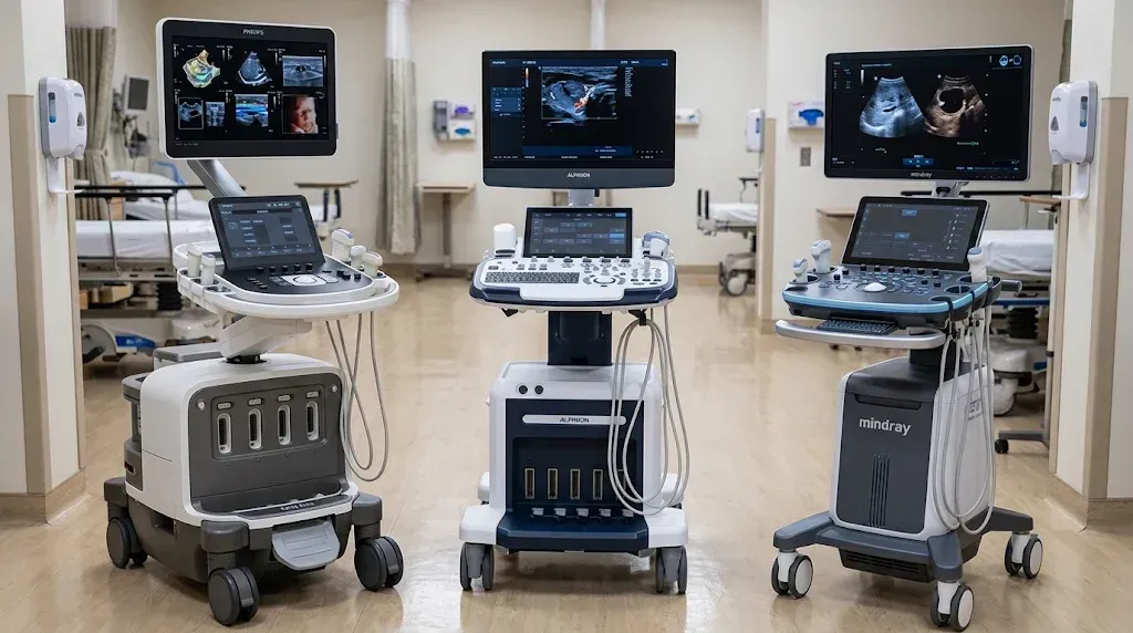
Decode Philips, Mindray, and Alpinion women's health ultrasound feature names, from TrueVue and iLive to Smart OB and X+ MicroView, and compare clearly.

Ultrasound buying glossary: demo vs. pre-owned, FMV vs. $1 buyout leases, service contracts, uptime, software licenses, and IT terms explained for practices.
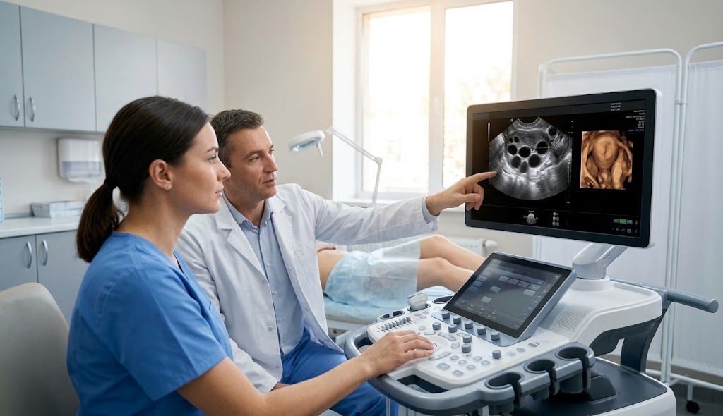
Choosing an ultrasound machine for an IVF or fertility clinic? Compare imaging, follicle monitoring, automation, workflow, and equipment costs.
Compare Philips, Mindray, and Alpinion OB/GYN ultrasound systems in 2026. Explore imaging technology, features, pricing, and the best fit ...
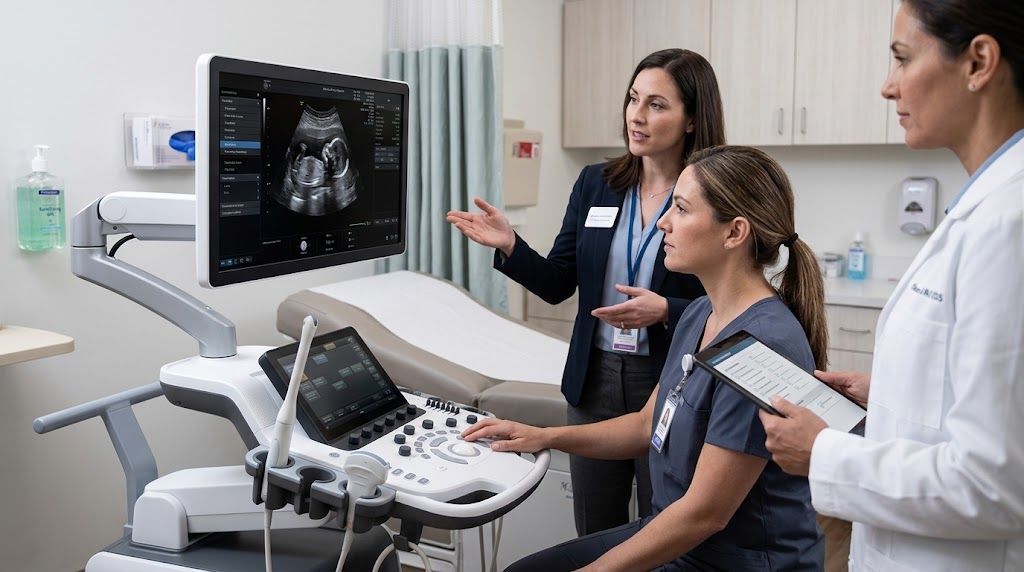
Learn how to evaluate an OB/GYN ultrasound machine demo, including image quality, probes, workflow, automation, ergonomics, and service.
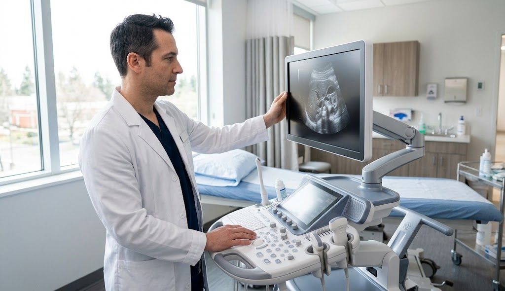
Explore Alpinion X-CUBE 90, X-CUBE 70 and X-CUBE i9 ultrasound systems for OB/GYN, fertility and women’s health imaging in 2026.
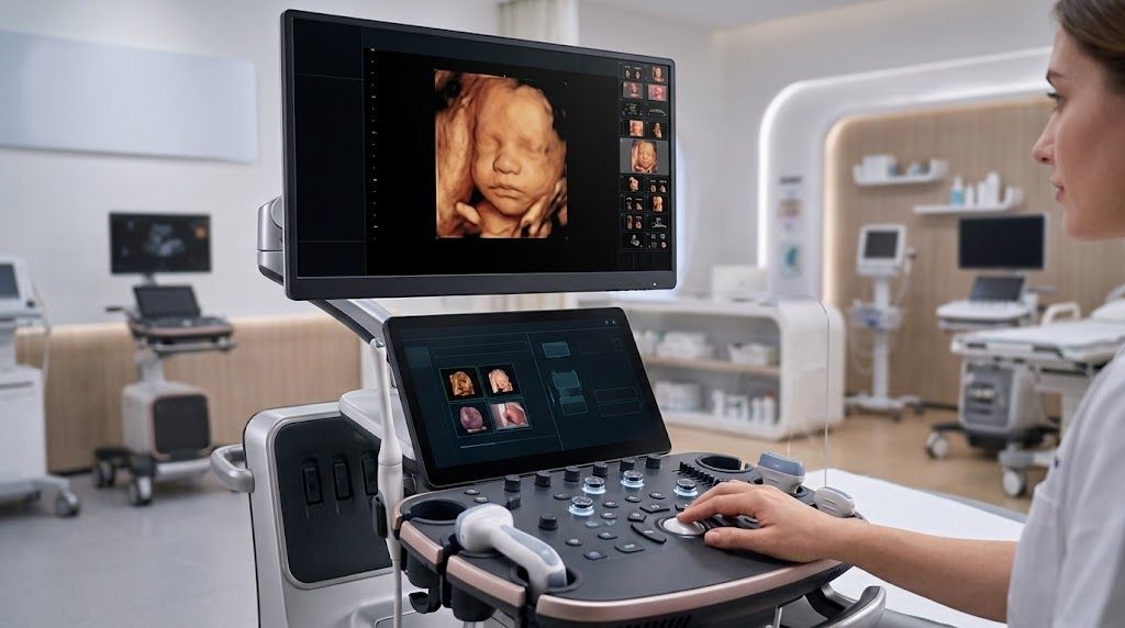
Compare Mindray Imagyn I9, Resona I9T and Consona N9 ultrasound systems for OB/GYN, fertility and women’s health imaging in 2026.
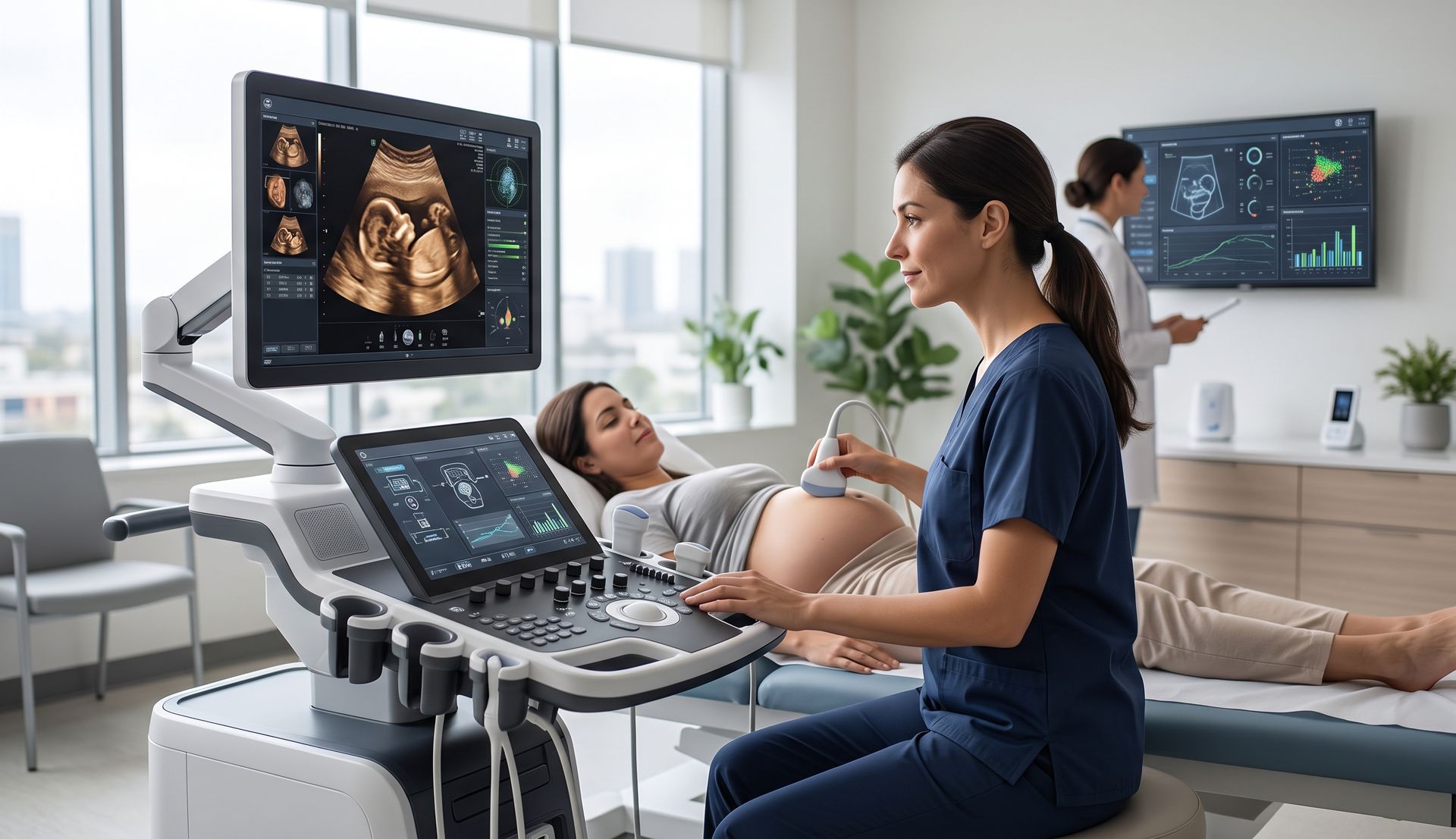
Explore Philips women’s health ultrasound in 2026, including EPIQ Elite, Affiniti 70 and Elevate Plus advancements in imaging, AI and workflow.
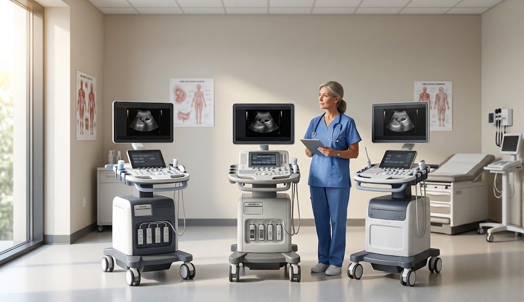
Compare a new vs refurbished ultrasound machine for OB/GYN use, including condition, warranty, software, probes, service support, and lifecycle value.

Should You Buy, Finance, or Lease an OB/GYN Ultrasound Machine in 2026? A Practice Acquisition Guide
Compare buying, financing, and leasing options for OB/GYN ultrasound equipment and learn which acquisition strategy best fits your practice.